Research
Three-dimensional biological imaging with a crystal interferometer
The Bonse-Hart-type X-ray interferometer shown in Fig.1 is made of almost-perfect silicon crystal, and is a historical interferometer for hard X-rays. The Bragg diffraction with the crystalline lattice divides and adds up the X-ray beam as shown in the figure, composing a Mach-Zehnder interferometer. The observation of the objects with weak absorption such as soft tissues and soft matters is possible with this type of interferometer as well (Fig.2). Because it involoves the interference of the two beams, the phase shift can be detected directly, and the highest sensitivity is possible with this method. We have worked on collaborative projects with medical doctors of Kobe University and the University of Tsukuba (an example is shown in Fig. 3).
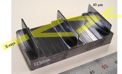
Fig. 1: the Bonse-Hart-type X-ray interferometer made of silicon. The yellow lines show the paths along which the X-ray propagates. To achieve high-resolution, the crystal plate on the right-hand edge is only 40 microns thick.
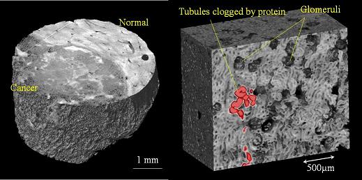
Fig.2: CT images obtained with this method: part of a rabbit liver (left) and part of a rat kidney (right).
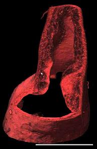
Fig. 3*: Three-dimensional rendering of the right common carotid artery of the ApoE-KO mouse that was fed a high-cholesterol diet. Scale bar 1 mm.
*Reproduced figure with permission from M. Shinohara et al., Am. J. Physiol. Heart Circ. Physiol. 294 (2008) 1094. Copyright 2008 by the American Physiological Society.