Development and application of X-ray phase microscopy
Grating-based X-ray phase imaging that functions with a spherical wave shows an excellent compatibility with X-ray microscopy. The spatial resolution attainable by a Talbot interferometer is limited by the period of gratings (several microns) because it is based on the moiré formation. Therefore, structures smaller than the grating period is not resolved. To overcome this limit, we are studying the combination with X-ray microscopy that can magnify sample images. Specific approaches include projection microscopy and imaging microscopy. Here, the latter is introduced.
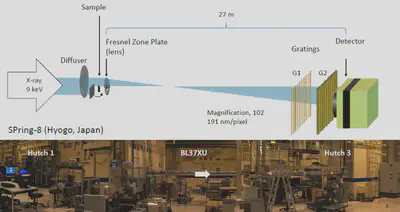
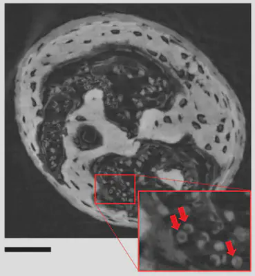
Fresnel zone plate (FZP) that has a function as an X-ray lens is used in X-ray imaging microscopes. We combined a Talbot interferometer and a FZP at SPring-8 (Fig. 1), and a spatial resolution of sub-µm in phase tomography has been achieved. In collaboration with Prof. K. Matsuo with Keio Univ., various mouse-bone samples are scanned to contribute to bone science (Fig. 2) [1].
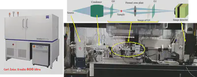
In our laboratory, a laboratory-type X-ray imaging microscope (Zeiss Xradia 800 Ultra) is in operation, and we are developing an X-ray phase microscope by combining gratings. Although the X-ray tube used in the microscope does not ensure the spatial coherency, the confabulation of so-called Lau interferometer [2] was introduced, and the mode of phase imaging/tomography has been successfully appended under a spatial resolution of 50 nm [3, 4].
The X-ray microscope is registered as a common apparatus of Tohoku University.
https://ses.tsc.tohoku.ac.jp/eq/front.php?cont=eq_detail&id=488 (university users)
https://ses.tsc.tohoku.ac.jp/public_eq/front.php?cont=eq_detail&id=488 (others)
-
[1] H. Takano et al., AIP Adv. 10 (2020) 095115. DOI:10.1063/5.0016318 ↩︎
-
[2] A. Momose et al., Appl. Phys. Express 4 (2011) 066603. DOI: 10.1143/APEX.4.066603 ↩︎
-
[3] H. Takano et al., Appl. Phys. Lett. 113 (2018) 063105. DOI: 10.1063/1.5039676 ↩︎
-
[4] H. Takano et al., Optica 6 (2019) 1012-2015. DOI:10.1364/OPTICA.6.001012 ↩︎